contact
Jade Sutton
Administrative Assistant II
AMED: Academy of Microscope Enhanced Dentistry
13th Annual Meeting and Scientific Session: "Pathways to Perfection"
Friday, Nov. 14 through Sunday, Nov. 16 at the University of Maryland, Baltimore's Southern Management Corporation Campus Center, 621 W. Lombard St., Baltimore, MD 21201
Hands-on courses at the University of Maryland School of Dentistry, 650 W. Baltimore St., Baltimore, MD 21201
Friday, Nov. 14
Clinical Benefits of a Microsurgical Approach in Periodontal and Peri-Implant Surgery - New Insights Into Biology of Wound Healing
Presented by: Rino Burkhardt, DDS, DMD
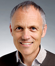 Speaker Biography:
Rino Burkhardt graduated from the University of Zurich and received his doctorate from the Medical Faculty of the same University. He is an EFP (European Federation of Periodontology) certified specialist in periodontology and received his Masters degree from the Medical Faculty of the University of Berne (MAS in Periodontology).
He maintains a private practice in Zurich, limited to periodontology and implantology. He's an active member of the European Academy of Esthetic Dentistry (EAED), the European Association for Osseointegration (EAO), the Swiss Society of Periodontology (SSP) and Board member of the Swiss Society of Implantology (SGI).
Course Description:
Periodontal plastic surgery has developed in the last 25 years and is indicated to solve functional and/or esthetic problems of the hard and soft tissues of the oral cavity. A considerable amount of these interventions are technically very sensitive and therefore, the results may vary a lot. Well investigated are the regenerative procedures and the coverage of mucosal dehiscences. The interest of the researchers has mainly been focused on two aspects, namely on the regenerative materials and products on one side and on novel surgical approaches and improvements on the other side. Concerning the latter, it has been shown in other surgical specialties that the use of a surgical microscope can enhance the clinical outcome and minimize the morbidity of the patients. Nevertheless, in periodontal and peri-implant surgery, the use of the microscope never established like in ophthalmology or vessel surgery and the single variables which are responsible for the improved results have never been elucidated in detail.
It is the aim of this lecture to present the indications for the use of a surgical microscope in periodontal and peri-implant surgery and to carefully evaluate why there is a benefit for the patients when treated with a minimally-invasive technique. Illustrated by clinical cases, the differences between a minimally-invasive approach and a conventional approach will be shown. Additionally, a new deeper insight into the biology of wound healing has shown the potential mechanisms which are important for the better clinical outcomes.
New Frontiers in Periodontal and Bone Regeneration
Presented by: Mark A. Reynolds, DDS, PhD, MA
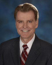 Speaker Biography:
Dr. Reynolds is professor, Dean and chair of the Department of Periodontics at the University of Maryland School of Dentistry. He holds a master's degree in clinical psychology, a specialty certificate in periodontics, and a PhD in oral and experimental pathology. He is a Diplomate of the American Board of Periodontology and holds fellowship in both the American College of Dentists, International College of Dentists, and Pierre Fauchard Academy. Dr. Reynolds is currently a Director of the American Board of Periodontology. He has served as a consultant to research and regulatory agencies, including the NIH and FDA, and serves on the editorial boards of four scientific journals. He has contributed over 125 clinical and scientific peer-reviewed publications, and his interests include inflammation, periodontal and bone regeneration, and dental implants.
Course Description:
An understanding of regenerative techniques and materials is critical for achieving successful and predictable bone and periodontal regeneration in clinical practice. This program will review current therapeutic techniques and materials as well as emerging technologies, including stem cells and lasers.
p
p
Dental-Labial Harmony through Cosmetic Dentistry and Injectables
Presented by: Laurence Rifkin, DDS
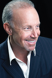 Speaker Biography:
Dr. Laurence R. Rifkin practices dentistry in Beverly Hills, California, with a focus on Dento-Facial Aesthetics and Comprehensive Periodontal-Implant Rehabilitation. Dr. Rifkin graduated USC School of Dentistry in 1976 where he received his dental degree.
As an international lecturer, Dr. Rifkin has spoken to numerous dental and cosmetic surgery academies on various topics such as: Team Approach to Comprehensive Facial Beauty, Natural Beauty for Teeth and Implants, Microscopic Dentistry, and Complete Mouth Rehabilitation.
In addition to lecturing, he has also been a faculty member of both USC and UCLA’s schools of dentistry. Dr. Rifkin is also a member of the Academy of Microscope Enhanced Dentistry, American and European Academies of Esthetic Dentistry as well as one of the few dental members of the American Academy of Cosmetic Surgery. He has published numerous papers in Facial Aesthetics and restorative and implant supported dentistry.
Dr. Rifkin is passionate about combining a foundation of excellence in function, periodontal health and Dento-Facial aesthetics utilizing science, technology, and fine arts. He believes in a team approach to achieving the optimal result for our patients working with both dental and medical specialists in both health and Facial Cosmetics.
Course Description:
Contextualism in architecture and art is: "The aesthetic position that a building or the like should be designed for harmony or a meaningful relationship with other such elements already existing in the vicinity."
This ancient Greek philosophy holds true in Aesthetic Dentistry as well. The execution of this ultimate end goal is built upon the observation of these elements both micro and macroscopically. We must have the combined vision and skills to achieve this outcome. Hence, we as dental professionals must acquire the control of both hard and soft tissues that interact harmoniously creating a beautiful smile and face. This presentation will address the diagnosis and treatment of the soft tissue contours of the surrounding lower face and lip structure to complete the dento-gingival complex. In addition to aesthetic dentistry, the utilization of neurotoxins and dermal fillers as injectable solutions to the aging face, soft tissue asymmetries and soft tissue augmentation will be described with techniques and before and after examples will be presented.
Microsonic Management of Calcified Canals
Presented by: Noushad Rahim, BDS, MDS, MFGDP, MJDF RCS Eng
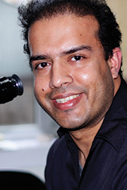 Speaker Biography:
Noushad is an endodontist practicing in the United Kingdom. He obtained his MDS in Endodontics and Conservative Dentistry from Manipal University (India) in 1998. He worked for more than 7 years as an Endodontist in the United Arab Emirates and was the Course Coordinator and Internal Examiner for Clinical Endodontics at the College of Dentistry, Ajman University. He has also lectured and conducted hands on workshops both nationally and internationally. He is an opinion leader and lectures for some of the leading UK endodontic dental suppliers. He is a member of the British Endodontic Society. He has been voted into the Elite 20 by readers of Private Dentistry Magazine, UK (2013). Outside dentistry, Noushad is a keen supporter of Liverpool "Soccer" Club and enjoys traveling with his wife and 2 daughters.
Course Description:
Locating and negotiating a calcified canal for non-surgical root canal therapy can be both taxing and frustrating to the dental practitioner. The prognosis of a tooth indicated for endodontic therapy can rapidly deteriorate during attempts to find the calcified canal. Risks of perforation, blockages, and instrument separation are high in these challenging cases. The use of an operating microscope and ultrasonic has made locating the calcified canal more predictable and safe. This presentation will outline approaches to manage calcified canals.
Protocol of Preparation for Full Crowns and Veneers with Microscope - Full Mouth Micro Invasive Rehabilitation
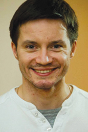
Presented by: Nazariy Mykhaylyuk, DMD
 Speaker Biography:
Dr. Mykhaylyuk graduated from Ivano-Frankivsk Dental Academy (Ukraine) in 2008. He specializes in micro dentistry and practices at his family practice, "Dental Clinic of Mykhaylyuk - Oral Design Center Ukraine". Dr. Mykhaylyuk is a member of Rosmicro group, ITI, AMED, ACE and is the Co-founder of MicroVision Group.
Course Description:
Nowadays technologies in dentistry are improving very fast. But whatever technology or approach you use, you are supposed to visually control all your steps. So when it comes to prosthetic dentistry (analysis, preparation, provisionals restoration, impression control), magnification is really important to get the result we want in the end. We'll discuss the use of microscope in prosthetics on every step of the patient's treatment. We will implement the microscopic approach into a full mouth rehabilitation—macro and micro together.
Ultrasonic Preparations: Myth, Magic, and Magnification
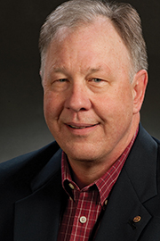
Presented by: Jeff Hamilton, DDS
 Speaker Biography:
Graduate of the University of Washington School of Dentistry in 1976, followed by a Certificate of Completion, General Practice Residency at Hennepin County Medical Center (University of Minnesota) in 1977.
Current professional affiliations include: AMED, Associate member of American Academy of Periodontolgy, ADA (and state & local affiliates), Pacific Coast Society for Prosthodontics, and American Academy of Oral Maxillofacial Radiology (associate).
Current academic affiliations: University of Washington Dept. of Oral Medicine (Affiliate Faculty) and UCLA Restorative Dept. (Lecturer).
Maintains a private general dental practice in Olympia, WA and is a lifetime student of general dentistry and patient care with dental laboratory microscope experience since 1982 and clinical microscope since 2000.
Out of the office life includes wife, Lorraine, and family of five (ages 17-31). Also an active member in the National Ski Patrol (White Pass, WA) for 24 years teaching outdoor emergency care, avalanche, and mountain travel.
Course Description:
In 2005, Dr. Domenico Massironi introduced many of us attending AMED to ultrasonic precision crown margin finishing. This led to my own journey in studying ultrasonic power sources (handpieces), diamond tip coatings, tip design, effective use, and product evaluations. This summary of findings and clinical examples of ultrasonics will review uses in restorative, periodontics, and endodontics.
Microscopically Guided External Sinus Floor Elevation (MGES) - A New Microsurgical Protocol in Oral Implantology
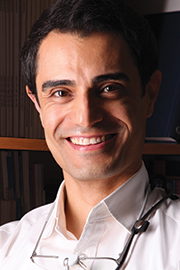
Presented by: Behnam Shakibaie, Dr. med. dent
 Speaker Biography:
- Studies in Dentistry at Berlin University 1993-1998
- Doctorate at Berlin University due to clinical Guided Bone Regeneration (GBR) 1996-2001
- Specialization in Oral Surgery 1999-2003
- Mastership Implantology & Periodontology (DGI/DGP) 2004-2007
- Specialist in Oral Micro Surgery & Micro Dentistry of Carl Zeiss Academy 2007-2009
- Specialist of Oral and Maxillofacial Surgery in Iran 2007
- Specialized clinic in minimally invasive, microscopically guided Implantology & Periodontology in Rheda-Wiedenbrueck/Germany 2005-2009
- Since 2011 Specialized clinic for microsurgical Implantology & Periodontology in Tehran/Iran
- Since 2005 Invention, development and publication of microsurgical operation techniques and instruments in oral implantology
- Scientific Consultant of DGI (German Association of Implantology)
- Instructor of Department of Implantology of Carl Zeiss-Dental Academy
- Ambassador of Quintessence Publishing Group
Course Description:
Sinuslift procedure has become the most scientifically recognized bone augmentation technique in posterior maxilla in oral implantology. But intraoperative complications such as sinus membrane perforations, the high rate of patient morbidity related to the extensive surgical trauma and insufficient implant primary stability still cause surgeon's and patient's discomfort. Therefore in recent years intraalveolar (internal) sinuslift is getting more popular. But especially in cases with advanced 3-dimensional alveolar bone atrophy internal sinuslift is contraindicated. Therefore Shakibaie published the new surgical technique of MGES (Microscopically guided external sinus floor elevation) in 2008 and 2010 as a minimally traumatic alternative to internal sinuslift. MGES enables the microsurgically experienced surgeon to reduce the trauma of sinuslift significantly and to increase the safety of this technique too. MGES main conditions are optical magnification of surgical microscope or loupe, specially developed microsurgical sinuslift instruments, technically trained surgeon and assistance.
Saturday, Nov. 15
Minimally Invasive Interventions for Esthetic Dentistry
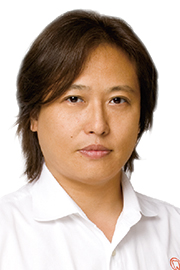
Presented by: Masayuki Okawa, DDS
 Speaker Biography:
Dr. Masayuki Okawa graduated and received his DDS from Touhoku Dental University, School of Dentistry in 1986. He maintains his private clinic in Tokyo from 2001. He is a director of SJCD Tokyo, a certified dentist of the Japan academy of occlusion and an active member of the Japan academy of esthetic dentistry, etc.
Course Description:
Lately, favorable results are seen in many esthetic cases with minimally invasive techniques. This became possible due to the development of biomimetics, advancement in bonding technique, and treatment using the problem-based approach. In addition, the use of the microscope has allowed us to obtain precise and predictable outcomes.
Using my Minimally Invasive Full Mouth Rehabilitation cases, the use and effectiveness of the microscope in direct composite and porcelain-bonded restorations in esthetic dentistry will be discussed.
Microscope Enhanced Restorative Dentistry: A Prosthodontic Perspective
Presented by: Keith Boenning, DDS
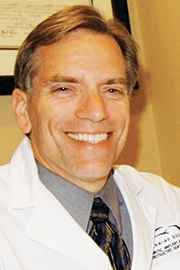 Speaker Biography:
Dr. Keith A. Boenning specializes in Prosthodontics. His focus is with cosmetic, implant, and reconstructive dentistry and he has been practicing full time in Baltimore since 1986. After graduation from the University of Maryland School of Dentistry in 1983 , he stayed and completed his advanced specialty training in Prosthodontics in 1985.
A firm believer in the importance of sharing professional expertise with colleagues, Dr. Boenning participates in various study groups and lectures extensively on dental procedures, innovations in the field of cosmetic dentistry and dental implants. He teaches in two post-graduate programs at the University of Maryland: Advanced General Dentistry and the Implant Periodontal Prosthodontics Program.
Dr. Boenning is a member of the American College of Prosthodontics, the Academy of Osseointegration, the American Academy of Esthetic Dentistry, the American Academy Of Cosmetic Dentistry, the American Dental Association, the Maryland State Dental Association and the Baltimore County Dental Society. Dr. Boenning is also a Board Member for the American Heart Association in Baltimore.
Course Description:
I use the scope 100% of the time for restorative procedures. I will explain why I transitioned from just working on maxillary anteriors and margin refinement to using the microscope for all procedures, and how I did this. Then I will cover the four sitting positions I use for access to any tooth and tooth surface, packing cord, impression techniques, surgical benefits and many other exacting procedures that allow me to create accurate and precise restorations.
Treatment of Newborns and Infants with Breastfeeding Difficulties - Using Lasers to Correct Tongue and Lip Ties
Presented by: Lawrence Kotlow , DDS, P.C.
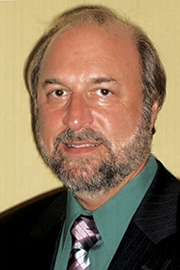 Speaker Biography:
Dr. Lawrence Kotlow is a 1972 graduate of SUNY Buffalo Dental School, and completed his pediatric dental residency at the Children's Hospital in Cincinnati, Ohio between 1972-1974. Since 1974 he has had a private practice in Albany, New York. He became Board Certified in Pediatric dentistry in 1980, and is a Fellow in the American Board of Pediatric Dentistry. Dr. Kotlow has received the three major Third District Dental Society Awards; he has served as President of the Third District Dental Society of New York State and served on many committees at the State level. He is a member of the ADA, ICD, New York State Dental Association, as well as a member since 2000 of the ALD. He is a founding member of the International Affiliation of Tongue-tie professionals, a group dedicated to improving infants and mothers ability to breastfeed.
Course Description:
This introduction to infant diagnosis, treatment and post surgical care will describe the three primary lasers used for soft tissue surgery: the 1064 diode, the Erbium:YAG and the CO2 laser (9300nm). The classification, correct method for evaluation and diagnosis of infants and post surgical care will be presented.
This presentation will provide the educational materials and practical information for participants to understand the way to treat and diagnose oral tongue and maxillary lip tie problems in newborns, infants and young children.
Papilla Reconstruction Around Teeth and Implant Using Connective Tissue Graft
Presented by: Junya Okawara, DDS
 Speaker Biography:
AMED member 1994: Graduated from Nihon University School of Dentistry at Matsudo. 1998: Completed Postgraduate Studies at Nihon University. 1999: Worked with Dr Masana Suzuki at the Suzuki Dental Clinic. 2003: Opened Private Dental Clinic in Tsukuba City, Japan.
Course Description:
Inter-dental and inter-implant papilla defects in the anterior region can be a major esthetic concern for some patients. However, it is very difficult to reconstruct dental papilla defects in the anterior region. Although the techniques of papilla reconstruction around teeth and implants have been described, none of the currently available surgical procedures for dental papilla reconstruction provide predictable results.
Recently, the relationship between papillae height and facio-lingual thickness of the papilla base have been described. In other words, if we want to increase the height of the dental papilla, that thickness also must be increased. This time, I will make a presentation on dental papilla reconstruction using a connective tissue graft around the teeth and implant. And I will talk about the importance of the soft tissue thickness of the papilla base for dental papilla reconstruction.
Tooth Anatomy, Endodontic File Design and Dentin Conservation - A New Paradigm
Presented by: Eric Herbranson, DDS
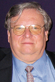 Speaker Biography:
Dr. Eric Herbranson is co-founder and Executive Director of Brown & Herbranson Imaging, a company that develops dental and human anatomy education software. With over 30 years in practice, Dr. Herbranson is a dedicated clinical endodontist. He has made a significant contribution over the last 20 years as clinical assistant professor at the University of the Pacific School of Dentistry where he lectures to students and special interest groups on endodontics, technology in dentistry, and microscope photography. Dr. Herbranson s study of physics and 40 years experience in film and digital imaging provide him with an educated understanding of macro and microphotography, and affords him a unique vision of endodontic education and image production. With his innovative approach and advanced imaging skills, Dr. Herbranson developed the unique processes and methodology for capturing images of human and dental anatomy now used as the basis for Brown & Herbranson Imaging s educational technology. Dr. Herbranson is the co-author of the chapter on tooth anatomy in Pathways of the Pulp, the topselling textbook on endodontics. Dr. Herbranson is a frequent speaker and educator at universities and conferences on the subjects of integration of new technology into dentistry, the use of software and computers in presentations, and surgical operation microscope photography. He is actively involved as a consultant in exotic animal dentistry for the Oakland Zoo. Dr. Herbranson earned a Bachelor of Science from La Sierra College, a Doctoral of Dental Surgery from Loma Linda University,and a Masters of Science in Endodontics from Loma Linda University.
Course Description:
This lecture will review how a nuanced understanding of tooth anatomy and an appreciation of the value of conserving dentin will change endodontic access and shaping protocols. It will explore the impact of endodontic file design on restorative failure in endodontically treated teeth. It will also discuss the importance of conservation of pericervical dentine as a factor in preventing restorative failure of endodontically treated teeth. It will present new instrument design that automatically and safely create more root form appropriate shapes. And it will outline an approach to endodontic access design that feature dentin conservation as an objective.
Intervention Study About the Effect of Arm Supports on Muscle Activity, Posture and (Dis)comfort in the Neck and Shoulder Region in Microscope Dentistry
Presented by: Jacqueline Bos
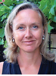 Speaker Biography:
Jacqueline Bos, physical therapist and ergonomist, established BBO in 2005. BBO provides ergonomic evaluation and training in the workplace and assists in designing or adapting workplaces. BBO is specialized in dental ergonomics, veterinary ergonomics, and ergonomics for medical specialists. Jacqueline lectures (inter)nationally about dental ergonomics and the specific aspects of dental specialties such as endodontics and implantology. Jacqueline's mission is to teach people to work in a healthy manner. Thanks to her background as a physical therapist, she is able to give students real experience in working comfortably, safely, and efficiently.
Course Description:
Dentistry is considered to be a demanding profession because of the level of concentration and precision required for good performance. Dental work implies many precision tasks, with lots of visual challenges and demanding precision of movement, sometimes in combination with the application of heavy forces and psychosocial stress. In fact dentists would need some of Superman's superpowers to be able to perform optimally.
Multiple ergonomic solutions are designed to reduce the workload, without their effectiveness ever having been scientifically evaluated. A group of 11 endodontists was asked to perform a representative endodontic task under three conditions: 1) with Flexible arm supports, 2) with Fixed arm supports and 3) without arm supports. The self reported discomfort, posture observation and surface electromyography of neck and shoulder were assessed as outcome measures. In this lecture the results of this study will be shown: which situation will decrease the physical load? Challenge yourself and start taking measures to be able to work like Superman.
Sunday, Nov. 16 - Hands-On Courses
Endodontics: Conservative Access and Shaping Design
Presented by: Eric Herbranson, DDS
 Speaker Biography:
Dr. Eric Herbranson is co-founder and Executive Director of Brown & Herbranson Imaging, a company that develops dental and human anatomy education software. With over 30 years in practice, Dr. Herbranson is a dedicated clinical endodontist. He has made a significant contribution over the last 20 years as clinical assistant professor at the University of the Pacific School of Dentistry where he lectures to students and special interest groups on endodontics, technology in dentistry, and microscope photography. Dr. Herbranson s study of physics and 40 years experience in film and digital imaging provide him with an educated understanding of macro and microphotography, and affords him a unique vision of endodontic education and image production. With his innovative approach and advanced imaging skills, Dr. Herbranson developed the unique processes and methodology for capturing images of human and dental anatomy now used as the basis for Brown & Herbranson Imaging s educational technology. Dr. Herbranson is the co-author of the chapter on tooth anatomy in Pathways of the Pulp, the topselling textbook on endodontics. Dr. Herbranson is a frequent speaker and educator at universities and conferences on the subjects of integration of new technology into dentistry, the use of software and computers in presentations, and surgical operation microscope photography. He is actively involved as a consultant in exotic animal dentistry for the Oakland Zoo. Dr. Herbranson earned a Bachelor of Science from La Sierra College, a Doctoral of Dental Surgery from Loma Linda University,and a Masters of Science in Endodontics from Loma Linda University.
Course Description:
This workshop will allow the participant to understand and practice the newer concepts in dentine conservation in endodontic access and shaping. It will use plastic and extracted teeth along with newer design access burs and NiTi shaping burs to demonstrate and train the concepts.
Techniques of Micro Suturing
Presented by: Arnold Sindler, DDS
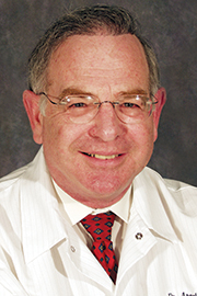 Speaker Biography:
- Bachelor of Science in Engineering from Johns Hopkins University —1966
- Dental Degree from University of Maryland School of Dentistry — 1976
- Certificate in Periodontology from University of Maryland School of Dentistry — 1978
- Clinical practice limited to Periodontics and Implant Dentistry in Westminster, Md.
- Associate Clinical Instructor -- University of Maryland Dept. of Periodontology
- Founding Member and Officer Gerald M. Bowers Study Club in Periodontology
- Member & former Board of Directors member Academy of Microscope Enhanced Dentistry
- Director Periodontal & Implant Microsurgery, University of Maryland Maryland School of Dentistry Department of Periodontics
Course Description:
This half-day program is structured to provide introductory information and hands on practice with micro-suturing. Instrument design, suture needle terminology, hand position, knot tying, and body support will be presented. Participants should have basic knowledge of operating the microscope.
Documentation with the Surgical Microscope: Improved Communication with Students, Colleagues and Patients
Presented by: Wayne Remington, DDS
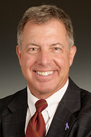 Speaker Biography:
Dr. Wayne Remington: A graduate of Temple University School of Dentistry in 1982 and the University of Virginia Medical Center General Practice Residency in 1984, Dr. Remington has been in private general and esthetic restorative dentistry since 1984. In addition, he has been a Clinical Assistant Professor of Dentistry at the University of Virginia, teaching esthetic dentistry.
Dr. Remington has been using a microscope in restorative practice since 1997 and been teaching two-day hands-on courses since 1999. His courses are designed to teach a maximum of four individuals at a time for personalized attention.
Course Description:
This course is a live demonstration of the how's and why's of connecting video and still cameras to surgical microscopes. The ability of demonstrating what is going on at the business end of our hand piece connects staff with the procedure at hand and keeps them engaged in the process. The same ability gives unmatched communication to the patient on the procedure being performed. And most importantly for educators, this ability allows the instructor to demonstrate clearly to ALL the students (not just the ones standing next to your shoulder) exactly what is going on at the tip end of your bur!
Many dentists and dental educators have had difficulty getting easily obtained images for instructional purposes. This course will show you in detail how to connect cameras via HDMI or other cables to video outputs or computers; also we will demonstrate wireless technology for NFC (near field connectivity) or WiFi connections to tablets or smartphones. Participants are invited to bring their current camera systems with them (including beam splitter and mounting arm) if they wish personal coaching on their particular camera system. Please give information about your system to AMED prior to the course so that we may be adequately prepared to answer any questions about your system.
Tuiton:
- General registration with special UMSOD discount $595 (regularly $895)
- Full-time UMSOD faculty: $295 (regularly $895)
- UMSOD student: $195 (regularly $245)
- Use the discount code UMSOD at checkout
CDE Credits:
- Earn up to 16 CE credits
