


Research Technologies
Magnetic Resonance Imaging (MRI)
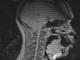
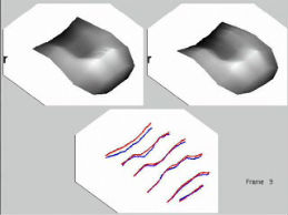
Vocal tract images are made in multiple planes during sustained speech sounds. Tagged Cine-MRI (tMRI) allows tracking of tissue points in oral structures.
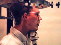
Electropalatography (EPG)
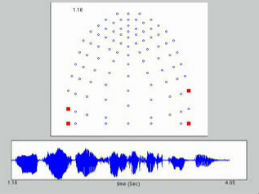
Acrylic palates, custom made to fit each subject's palate, record tongue-palate contact in real time.
Research Areas
Modeling the Tongue
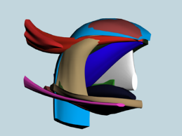
Modeling the interdigitated tongue muscles tests the many muscle combinations used to produce speech.
Tongue Segmentation
The tongue can be segmented into regions that produce a speech gesture using coordinated muscle activation strategies.
Swallowing
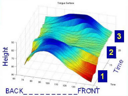
Tongue surface deformation creates a transverse waveform that propels the bolus from the oral cavity (front) into the pharynx (back).
Glossectomy Study
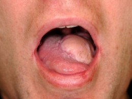
We study speech adaptation to glossectomy surgery, which removes a malignant tumor from the tongue. A ‘flap’ and may be used to replace the tissue.
Geometry to Acoustics
.jpg)
Vocal tract airway sections are calculated from reconstructed 3D super-resolution volumes, and used to generate speech spectra.