


Glossectomy Study
"Predictors of Speech Quality After Tongue Cancer Surgery"
Funded by NIH grants R01 - CA133015 and R01DC014707
Aim 1. Assess the degree to which internal tongue motion explains the relationship between speech quality and surgical procedure.
Develop an atlas of normal motion in 2D and 3D using Tagged MRI Data. Use Principal Components Analysis to compare these patterns to speakers who have had tongue cancer surgery.
Velocity fields show motion from /s/ to /u/
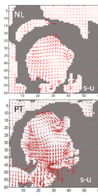
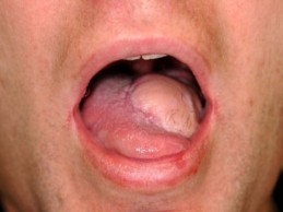
Note anterior depression behind tongue tip in patient. The patient has a lateral lisp.
Aim 2: Assess the degree to which muscle mechanics explains the relationship between speech quality and surgical procedure.
Track motion of muscles using Diffusion Tensor Imaging (DTI) to identify muscles and Tagged MRI Data to track them. Compare muscle mechanics of speakers after tongue cancer surgery to control speakers.
DTI fiber tracking shows IL fiber directions
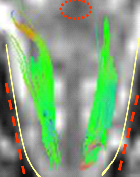
Control
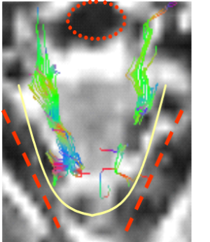
Patient
Axial View
Fiber directions:
Green: anterior-posterior
Blue: superior-inferior
Red: lateral-media
Tongue is outlined in yellow
Pharynx is circled in red
Mandible is tracked in red
In-plane Rotation and Elongation of Averaged Fibers
| "asouk" | Rotation | Rotation | Elongation | Elongation |
|---|---|---|---|---|
| schwa | 0 | 0 | 0 | 0 |
| start /s/ | 0.5 | -5.3 | 5.1* | 18.9* |
| max /s/ | -1.3 | -9.1 | 0.5 | 17.3 |
| /u/ | -20.2 | -4.0 | -2.7+ | 7.0+ |
| /k/ palate | -18.1 | -3.3 | -9.7+ | 3.3+ |
| /k/ velum | -19.0 | -1.6 | -10.3+ | 5.0+ |
|
* The IL lengthened during /s/. PT had more elongation during the /s/. + Both subjects then shortened the fiber moving into /u/ and /k/. PT never returned to resting length. |
||||
This kind of analysis allows us to compare across different levels of the speech production process.
Aim 3: Develop a tool to estimate speech improvement from surgical alternatives.
Develop 3D FEM models of the tongue and airway for control and patient speakers to determine the effect of vocal tract modifications on speech.
Subjects
Control subjects. Patients who have had surgery to remove T1N0M0 or T2N0M0 tumors, with no chemotherapy or radiation therapy.
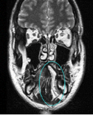
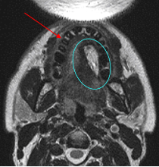
A radial forearm free flap replaced tissue removed in tongue cancer surgery
The flap appears white in the MRI because of its high fat content.
 |  |  |
|---|---|---|
| Sagittal | Coronal | Axial |