


Modeling the Tongue
The Visual Tract Vocalization Laboratory explores two models of the tongue: Representational and Predictive Modeling.
1. Representational Model
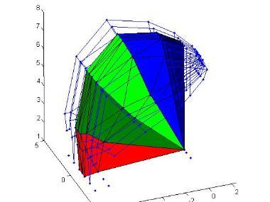
Principal Strains
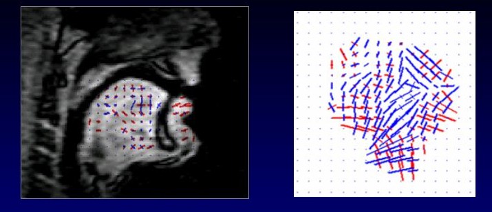 Blue = compression; Red = expansion |
At any location in the tongue, there are two orthogonal directions of primary stretch. Principal Strains show the amount of stretch in those two directions.
Movie of /i/-/u/

Midsagittal tMRI of /i/-/u/ (MICSR). The tongue moves back and down for /u/.
Movie of /a/-/u/

Midsagittal tMRI of /a/-/u/ with sound. The tongue moves up for /u/ and forward for the breath.
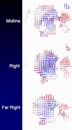
- The lower lip expands outward (red).
- The tongue tip compresses horizontally at midline, obliquely at sides.
- Hyoglossus compresssion appears at right.
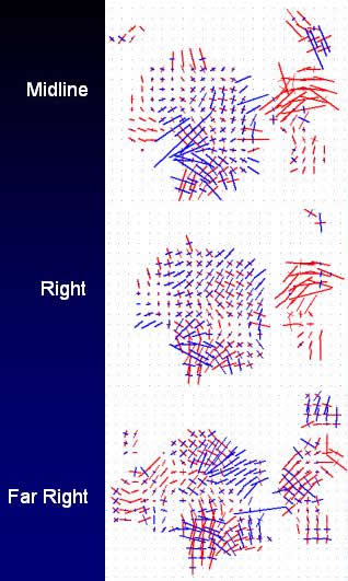
- The lower lip expands outward (red).
- The jaw muscles compress horizontally.
Predictive Model I
3D Finite Element Model of the tongue containing five muscles: genioglossus, transversus, verticalis, superior longitudinalis, inferior longitudinalis. Five segments of genioglossus are highlighted.
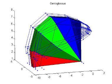
Undeformed
Front Raising
3. Predictive Model II
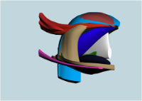
3D Finite Element Model of the tongue designed for Restore Medical Inc, Minneapolis, in conjuction with Reiner Wilhelms-Tricarico, Paul Buscemi, Mark Carlson, for the purpose of modeling sleep apnea. The model contains a jaw, hyoid and tongue with 3 extrinsic and 4 intrinsic tongue muscles (not palatoglossus); and three jaw/hyoid muscles (mylohyoid, geniohyoid, digastric).